Irreversible Electroporation (IRE), also known as NanoKnife, is a minimally invasive ablation therapy used in prostate cancer. It destroys prostate tumors using short electrical pulses rather than heat or radiation, allowing targeted destruction of prostate cancer cells while largely preserving surrounding structures such as nerves and blood vessels.
The use of IRE has broadened the therapeutic options available in prostate cancer. It allows defined tumour areas to be treated without removing the entire prostate, which has implications for functional outcomes and the ability to pursue additional treatments if needed.
IRE is primarily used as a focal treatment for localized prostate cancer but can also be applied in selected advanced or inoperable cases, depending on tumour extent and prior therapy.
This page explains the scientific principles behind irreversible electroporation, outlines how the NanoKnife procedure is performed, and reviews published clinical outcomes, recurrence patterns, and side-effect data in prostate cancer.
NanoKnife
A revolution in the treatment of prostate cancer
Irreversible Electroporation (IRE), also known as NanoKnife, is a minimally invasive ablation therapy used in prostate cancer. It destroys prostate tumors using short electrical pulses rather than heat or radiation, allowing targeted destruction of prostate cancer cells while largely preserving surrounding structures such as nerves and blood vessels.
The use of IRE has broadened the therapeutic options available in prostate cancer. It allows defined tumour areas to be treated without removing the entire prostate, which has implications for functional outcomes and the ability to pursue additional treatments if needed.
IRE is primarily used as a focal treatment for localized prostate cancer but can also be applied in selected advanced or inoperable cases, depending on tumour extent and prior therapy.
This page explains the scientific principles behind irreversible electroporation, outlines how the NanoKnife procedure is performed, and reviews published clinical outcomes, recurrence patterns, and side-effect data in prostate cancer.
Call us now on +49 (0)177 23 82 863 or get in touch using the contact form
Take The Time To Get Informed.
Prostate cancer often progresses slowly. In most cases, you will have enough time to learn about all treatment methods. In addition, classical “radical” therapy (surgery and/or radiation treatment) rarely prolongs life expectancy in low-aggressive prostate carcinomas – and in highly aggressive carcinomas, according to available statistics, it does not do so in many men. The same applies to radiation therapy.
At the same time, the classical treatments mainly lead to serious side effects, rarely to cure: 70-80% of all men experience erectile dysfunction after surgery, and about 20-50% of patients experience urinary incontinence: about 20% of men experience severe incontinence requiring several diapers per day, and an additional 30% experience moderate incontinence where one diaper per day is sufficient (see Table 1).
RT
3.2%
4.4%
9.4%
RT
60.8%
71.9%
93.9%
bowel function
RT
34.0%
31.3%
35.8%
Table 1: Long-term sequelae of radical prostatectomy (RPE) and radiotherapy (RT) for prostate cancer regarding incontinence, impotence, and impaired bowel function. Table translated into German. Resnik MJ, Koyoma T, Fan K-H, et al. N Engl J Med 2013; 368:436-45.
NanoKnife (IRE): Overview and Clinical Background
The NanoKnife tissue ablation procedure is a new type of therapy that is more than 10 years old and uses the irreversible electroporation (IRE) method to destroy cells. The surrounding tissue is not damaged in the process. Prostate cancer can be treated for the first time with this method in such a way that continence is preserved and there is only a low risk of impotence. In addition, the NanoKnife procedure has a very low likelihood of pain or scarring.
Here at VITUS, we are proud of the pioneering work we have done with the NanoKnife process. We are among the world’s leading experts in the use of IRE in the treatment of prostate cancer.
Through our close collaboration with the inventor of IRE for medical purposes, Prof. Boris Rubinsky of the University of Berkeley in the USA, we have had the opportunity to study and understand IRE at all stages of development, from laboratory to animal studies to human application. Our physicists and physicians are thus among the pioneers and world’s leading experts in electroporation processes – both in the scientific-technical field and in clinical use.
ADVANTAGES OF THE
NANOKNIFE TREATMENT
AT A GLANCE:
- One-time treatment completed in a single day
- Highest potency retention rate achieved to date*
- Statistical 0% incontinence**, ***
- No surgical incisions
- No aftercare necessary
- No higher recurrence rate than radical procedures**
- Treatment possible even after prostatectomy or radiotherapy
- Secondary immunological effects
* Urinary incontinence is the lack or inability of the body to safely store and self-directly empty the contents of the bladder. This leads to involuntary loss of urine. While there are several possible definitions of incontinence, a long-term study of incontinence after prostatectomy** concludes that a reasonable definition of incontinence is when a patient requires 2 or more pads per day 12 months after prostate surgery.
** Sacco E, Prayer-Galetti T, Pinto F, et al. Urinary incontinence after radical prostatectomy: incidence by definition, risk factors, and temporal trend in a large series with a long-term follow-up. BJU Int. 2006;97:1234-41.
*** Syan, Raveen, and Victor W. Nitti. “Post-prostatectomy Incontinence Initial Evaluation.” Urinary Dysfunction in Prostate Cancer. Springer, Cham, 2016. 15-30.
NanoKnife Technology for Prostate Cancer
The NanoKnife process is based on ultra-short pulses that generate strong electric fields. The pulses are 100 microseconds long – i.e. 0.0001 seconds. Compared to standard procedures, this new technology has unique features that make it ideal for treating the prostate.
- The NanoKnife process is based on ultra-short pulses that generate strong electric fields. The pulses are 100 microseconds long, i.e. 0.0001 seconds. Compared to standard procedures, thistechnology has unique features that make it well suited for treating the prostate.
- Tissue selectivity: Only cells that have a cell membrane, such as cancer cells, are reliably destroyed. All other structures, such as nerves, blood vessels and connective tissue structures, remain intact.
- Ultrasharp edges: With NanoKnife, the boundary between completely treated and untreated tissue can be measured in micrometers. This clearly distinguishes NanoKnife from other treatment methods, such as radiation therapy, heat-based treatments, and surgery. In these methods, the central treatment field is surrounded by an area in which tissue is unintentionally damaged, often extending several centimeters in diameter.
- Programmed cell death: NanoKnife induces cell death (apoptosis) without causing radiation damage or burns and without forming scars. Current standard methods, such as radiation therapy (proton therapy, brachytherapy, etc.) and heat-based therapies (HIFU, microwave therapy, etc.), create a large “toxic” area because the burned tissue is harmful to the body. This can result in inflammation, pain, and subsequent scarring, which may limit further treatment options in the event of recurrence.
- Repeatable any number of times: Because IRE treatment does not cause long-term tissue damage, other treatment methods (radiotherapy, heat-based treatments, or surgery) can still be performed after NanoKnife treatment. In addition, IRE treatment itself can be repeated as often as necessary, even after an initial irreversible electroporation treatment.
- Painless and minimally invasive therapy: No tissue is damaged by burning or radiation. Meanwhile, only thin needles are used as invasive instruments. As a result, patients usually notice little discomfort during treatment.
- Fast and in one session: Even extensive treatment areas can often be treated under general anesthesia in a single session.
- Wide range of possible applications: The treatment of small, early-detected prostate tumors is the simplest application of NanoKnife. However, NanoKnife can also be used to treat inoperable carcinomas that have breached the prostate capsule, as well as recurrences following radiation therapy, radical prostatectomy, high intensity focused ultrasound (HIFU) or brachytherapy.
- Immune system: Tumor fragments released during NanoKnife treatment support the patient’s immune system in combating further tumor growth.
Precision Requirements for NanoKnife Treatment
Since 2007, the IRE technique has been approved by the FDA in the USA and CE marking in Europe. This was done because corresponding studies prove that all cells within the treatment area can be killed with this method. In order to achieve this and carry out a successful IRE treatment, two factors are crucial:
- Firstly:
The exact position of the tumor must be known before treatment. - Secondly:
The treatment field must be placed in exactly the right position.
The first condition is fulfilled by multiparameter MRI examination, 3D biopsy and, if necessary, other procedures. The second condition is fulfilled by a minimally invasive procedure under general anesthesia. Sterile needles with variable exposure length are inserted. “Exposure length” refers to the area of the needle that exposes the tissue to electrical current. We have experience with this procedure since 2011 and have performed the most prostate treatments worldwide. This makes us one of the leading experts worldwide, while other clinics are just getting started.
Call us now on +49 (0)177 23 82 863 or get in touch using the contact form
The NanoKnife Treatment Procedure
This step is perhaps the most important. NanoKnife is a focal, image-guided treatment whose results are only as good as the planning and diagnostics that precede it. This is because NanoKnife itself does not detect where the cancer is located.
As a patient, you should not compromise on diagnostics. All variants of ultrasound (including elastography and contrast-enhanced ultrasound) as well as rectal punch biopsies have alarmingly low detection rates. It is very unlikely that this will find (all) tumors.
At the present time, only multiparameter MRIs performed by specialized radiology institutes provide adequate imaging diagnostics – together with PSMA/choline PET/CT if required.
The 3D images from MRI are given yet another layer by a 3D saturation biopsy. We are specialized in both procedures. Both are required to achieve the diagnostic accuracy necessary to effectively deliver NanoKnife treatment.
Once our physicians and physicists have determined the target area using the MRI cross-sectional images and, if necessary, calculated it in advance using 3D simulations developed by us, the target area is saved and transferred to the surgical monitor on the day of the operation.
For prostate treatment, the needles are inserted transperineally, through the perineum (the area between the genitals and anus). An endorectal ultrasound helps with placement. Experience plays a decisive role here. Even two needles place high demands on the spatial imagination of the surgeon. Placements with many needles are extremely challenging. This is because the prostate is a relatively small organ with a complex geometry, surrounded by many sensitive anatomical structures. In addition, the positioning options of the needles are very limited. This process can sometimes take several hours, although here at the VITUS Prostate Center computer programs help us achieve the perfect placement.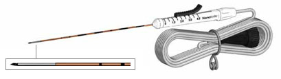
Figure 1: Minimally invasive NanoKnife electrode
Once the needles are precisely placed, their position is measured by ultrasound and passed on to the NanoKnife software and other external software. The area to be treated can still be adjusted on the computer by various parameters such as voltage, pulse duration and number of pulses. When all settings match the geometry of the prostate and tumor (always allowing for a safety margin around the tumor), NanoKnife charges its capacitors.
A potential difference builds up between each set consisting of two needles each. This is normally 3000 volts (V). This means that there is +1500 V on needle 1 and -1500 V on needle 2. The patient remains at 0 V. These voltages may seem more life-threatening than gentle, but their duration of action is in the range of millionths of a second. This makes the procedure safe and side effects such as burns are avoided. For more on the technology and the theory behind it, read more about the theory of IRE here.
The number of needles may vary and depends largely on the area to be treated. The computer of the NanoKnife system takes over the appropriate control of the needles, according to the previous settings. The treatment itself usually takes only seconds, a few minutes at most.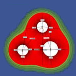
Figure 2: Precise planning of IRE treatment
Immediately after the needles are pulled out, the puncture sites close again. Most patients do not experience pain. However, some find the bladder catheter that is placed prior to treatment uncomfortable. It can be removed after 10 to 14 days. Small amounts of blood may still be found in the urine or seminal fluid several weeks later. This is because it takes up to six months for the body to move the tissue destroyed by NanoKnife ablation out of the body.
How IRE Compares With Radical Prostatectomy in Clinical Outcomes
While more than 2,000 men with prostate cancer have been successfully treated at the VITUS Private Clinic to date (as of January 2026), the published outcome data are based on 471 IRE treatments with a maximum observation period of 7 years².
Table 2 shows that the majority of the treated patients had high-risk prostate cancer according to the D’Amico classification.
Table 2
Most carcinomas were classified as stage T2a to T2c, meaning they were confined to the prostate and had not yet macroscopically penetrated the prostate capsule (“organ-confined disease”). However, 84 patients with T3 and T4 carcinomas were also treated, representing tumors that had already extended beyond the prostate capsule (“non-organ-confined disease”).
Our treatment outcomes show that IRE is not only suitable for the treatment of early, localized prostate cancer but also for the effective treatment of advanced carcinomas.
T4 N0 M1 /
T4 N1 M1
Table 3
During the observation period of up to 70 months, 4 recurrences occurred in patients with low-grade Gleason 6 carcinomas, 15 recurrences in patients with intermediate-grade Gleason 7 carcinomas, and 22 recurrences in patients with high-grade Gleason 8–10 carcinomas.
When compared with recurrence rates after radical prostatectomy (RPE), as reported in the Johns Hopkins University Han Tables, recurrence rates following IRE are comparable to those observed after RPE.
Recurrence Rates After IRE Treatment
How “Recurrence” Is Interpreted in Prostate Cancer
After radical removal of the prostate, one would expect the cancer to be completely removed from the body. However, this is not always the case and provides important context when interpreting recurrence rates after focal therapies such as IRE.
A closer look at the Han Tables shows that recurrence rates after radical prostatectomy remain substantial, even when the tumor was completely removed together with the prostate (the “organ-confined disease” category).
So where do these recurrences originate?
They arise from individual tumor cells that have migrated out of the prostate along connective tissue structures, lymphatic channels, and blood vessels, where they become embedded in the connective tissue surrounding the prostate. Tumor cells may also circulate in the bloodstream and can be detected using liquid biopsy techniques.
This occurs because the prostate capsule is permeable to cells, as it consists of compacted connective tissue fibers rather than a solid barrier. This can be visualized using the analogy of a forest edge: from a distance it appears solid, but on closer inspection it is largely open space with scattered structural elements.
Accordingly, many recurrences after prostatectomy occur in the connective tissue that previously surrounded the prostate and continues to surround the prostate region after prostate removal.
Strictly speaking, all local prostate cancer treatments represent tumor mass reduction, meaning the destruction or removal of tumor tissue within the prostate. Tumor cells located outside the prostate may remain in the body and, in successful cases, can be controlled by the patient’s own immune system.
Against this background, it is not surprising that recurrence rates after IRE treatment are comparable to those observed after radical prostatectomy and radiotherapy.
Observed Recurrence Rates After IRE Treatment
Table 4
Table 4 shows the number of recurrent prostate cancer tumors observed over a maximum follow-up period of 70 months in the different Gleason score groups among 471 IRE treatments performed at VITUS Private Clinic.
As expected, most recurrences occurred in patients with high-grade prostate cancer (Gleason 8–10), fewer in the Gleason 7 group, and the fewest in the Gleason 6 group.
Kaplan–Meier Analysis and Comparison With Radical Prostatectomy
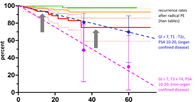
The graph in Figure 1 shows the Kaplan–Meier curves for the 471 IRE treatments evaluated. These curves illustrate the number of patients who developed prostate cancer recurrence over time following IRE treatment, stratified by tumor aggressiveness (Gleason 6, Gleason 7, and Gleason 8–10). The maximum observation period was 70 months.
Of particular interest is a comparison between recurrence rates in aggressive prostate carcinomas (red curve) and recurrence rates after radical prostatectomy (RPE), as reported in the Han Tables from Johns Hopkins University, USA (blue and pink dashed lines).
This comparison was performed using data from patients with comparable tumor stage and Gleason score.
Recurrence rates after IRE treatment in aggressive carcinomas fall within the “corridor” of recurrence rates observed after radical prostatectomy, meaning they are comparable.
Localization of Recurrences After IRE Treatment
The localization of recurrences provides additional insight.
Overall, only 8 of the 41 observed recurrences occurred within the IRE ablation field, and 15 occurred directly adjacent to the treated area. These cases can be attributed to surviving tumor cells located within or immediately outside the ablated tumor focus.
However, 16 of the 41 reported “recurrences” occurred in regions of the prostate that had been assessed as tumor-free prior to treatment and were therefore not treated.
These cases represent small, previously undetected carcinoma foci, most commonly in patients who declined comprehensive diagnostic evaluation, such as 3D mapping biopsy (3DMB), high-quality endorectal MRI, and/or PSMA-PET/CT with gallium-68.
In the strict sense, these cases do not represent true recurrences, but rather additional tumor foci that were not identified prior to treatment.
If these “pseudo-recurrences” are removed from the statistics and the Kaplan–Meier curves are recalculated based on actual recurrences, longer recurrence-free survival times are observed. This effect is shown in graph X, where the Kaplan–Meier curve for recurrence-free survival shifts upward (gray arrows).
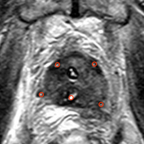
Clinical Example of Focal IRE Treatment

One ablation zone was generated per electrode pair (red ellipses). In the image after contrast administration, the ablation zone is clearly visible as an avascular area without blood supply. PSA levels decreased from 5.2 ng/ml before treatment to 2.3 ng/ml after IRE, rather than zero, because the right prostate lobe was preserved. This is typical of focal therapies.
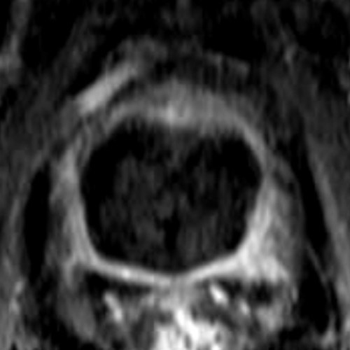
Figure 2 illustrates the treatment of a prostate carcinoma (stage T2b, N0, M0, Gleason 7b) located in the left dorsal outer zone (arrow; radiological orientation, view from below, right and left interchanged) in a 53-year-old man.
The treatment was performed using three electrodes, whose positions are visible on imaging the day after treatment (small red circles). One ablation zone was generated per electrode pair (red ellipses).
After contrast administration, the ablation zone is clearly visible as an avascular area without blood supply.
PSA levels decreased from 5.2 ng/ml before treatment to 2.3 ng/ml after IRE, rather than to zero, because the right prostate lobe was preserved. This is typical of prostate cancer focal therapies.
Safety Profile of IRE in Prostate Cancer
Table 5 below shows side effects that occurred during the treatment of 471 prostate cancer patients at the VITUS Private Clinic.
First, it is noteworthy that no life-threatening side effects and no treatment-related deaths occurred. This reflects the minimally invasive nature of the procedure.
Mild side effects occurred in approximately one-fifth of IRE-treated patients (green boxes). These included transient difficulty with urination (urinary retention), requiring reinsertion of a urinary catheter after removal of the initial bladder catheter.
In such cases, the catheter was typically left in place for an additional 10–14 days. Mild discomfort or pain during urination (dysuria) occurred in approximately 7% of patients, and mild blood in the urine (hematuria) in approximately 4%.
Moderately severe adverse events occurred in only 2.5% of IRE-treated patients (yellow boxes). These consisted of urinary tract infections related to catheter use, which were treated with antibiotics.
Medically relevant but non-life-threatening adverse events occurred in 1.3% of patients (pink boxes).
These included persistent urinary problems caused by necrotic tumor tissue within the prostate, requiring removal via the urethra (TURP); a fistula between the rectum and prostate caused by treatment, which healed spontaneously; and a bladder perforation caused by a urinary catheter, which also closed spontaneously.
Clinical or diagnostic monitoring only – no treatment required
(19.5%)
(9.0%)
(6.7%)
(3.8%)
Minimal, local or non-invasive treatment indicated; restriction of age-appropriate activities of daily living
(2.5%)
(2.5%)
(0.8%)
(0.8%)
(0.2%)
(0.2%)
Urgent medical measures required
Urinary Continence After IRE Treatment
At Vitus Private Clinic, we have experienced a 0% incontinence rate after IRE treatment in over 2,000 patients. By comparison, incontinence is a common and serious side effect in approximately 50% of men who have undergone radical prostatectomy, with or without da Vinci robotic assistance.
Incontinence is difficult to avoid during surgical removal of the prostate because the bladder sphincter, also called the lower urinary sphincter (LUS), is located directly below the prostate and extends into the apex, the lower part of the prostate.
If the prostate is removed, part of the sphincter is also removed. Additional damage to the LUS occurs when the base of the bladder is sutured downward after prostate removal and attached to the sphincter muscle to restore urine flow.
No incontinence has occurred in more than 2,000 patients treated with IRE to date. This was easy to ascertain because treated patients report even minor involuntary urine loss immediately. Nevertheless, immediately after IRE treatment, a small percentage of patients experience transient incontinence due to two mechanisms:
- Dilation of the sphincter muscle by the catheter, which remains in the urinary bladder for 10 to 14 days.
- Urge incontinence, which occurs due to irritation of the prostate gland after tumor destruction, similar to what is observed in prostatitis. This is noticeable when affected men experience a sudden urge to urinate when the bladder is full and may lose urine on the way to the toilet.
These forms of incontinence are temporary and typically resolve within a few days to a maximum of three months. They are not related to the irreversible destruction of the sphincter muscle that occurs after surgery.
Table 6 shows our published data on urinary incontinence.
Erectile Function After IRE Treatment
After IRE treatment for prostate cancer, impotence rates range from zero to a maximum of 10%.
The neurovascular bundle (NVB), a network of nerves and blood vessels, is responsible for the erectile function of the penis. It is located predominantly inside and directly adjacent to the prostate capsule (see Figure 2) and therefore cannot be preserved during either radical prostatectomy or radiotherapy.
As a result, impotence rates after conventional prostate cancer treatments are very high, ranging from 60–90%, even after so-called “nerve-sparing prostatectomy.” The outcome is loss of erectile function, i.e. impotence.

Clinical Examples: IRE in Advanced and Inoperable Prostate Cancer
IRE Treatment in Inoperable Prostate Cancer With Local Organ Infiltration
The treatment was performed with five electrodes, whose positions are visible the day after treatment (small black circles). One ablation zone was generated per electrode pair (red ellipses), resulting in a total of nine individual ablation zones. In the image after contrast administration, the total ablation zone is clearly visible as an avascular area without blood supply.
The prostate tissue was completely destroyed, while the prostate capsule was preserved, as it consists largely of connective tissue, which is not destroyed by IRE. PSA levels decreased to 0 ng/ml, comparable to levels after prostatectomy.
PSA 11 ng/ml
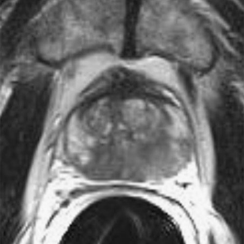
PSA 0.0 ng/ml
The images show treatment of a prostate carcinoma stage T2c, N0, M0, Gleason score 8, in a 63-year-old man. The carcinoma had spread across both lobes of the prostate. In this case, focal therapy with preservation of substantial portions of healthy prostate tissue was no longer possible.
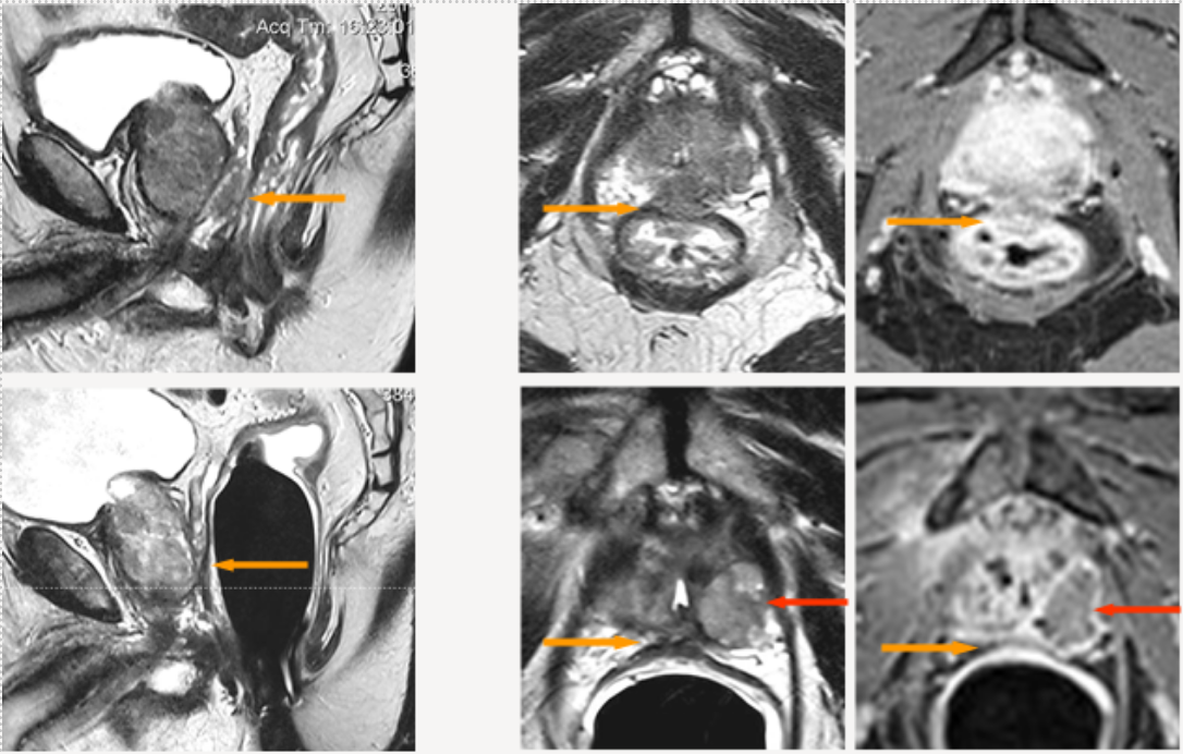
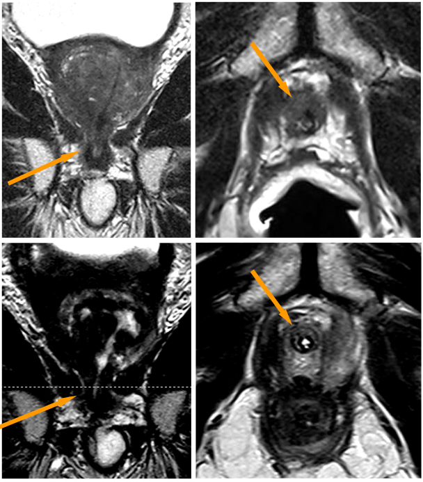
Complete Prostate Ablation Using Irreversible Electroporation (IRE)
Figure 3 shows treatment of a prostate carcinoma stage T4, N0, M0, Gleason score 8, in a 79-year-old man with infiltration of the rectum (rectal area indicated by yellow arrows). Radical prostatectomy would have required additional removal of the rectum and creation of permanent artificial bowel opening (anus praeter), requiring stool to be diverted into an external bag.
Rectal infiltration is shown in the MRI images in the top row of Figure 3:
- Left: sagittal image
- Middle: axial T2-weighted image
- Right: axial image after contrast administration
The lower row shows the findings nine months after IRE treatment. The rectal wall appears normal, without injury and without fistula (abnormal connection) to the prostate.
Inoperable Prostate Cancer and Advanced Treatment Options
At VITUS Private Clinic, we emphasize the importance of taking your time and finding the treatment option that is best suited to your individual case, starting with proper planning and accurate diagnostics. In some cases, standard diagnostic approaches do not fully identify inoperable prostate carcinomas before treatment decisions are made.
When the true extent of the tumor is only discovered intraoperatively, surgery may need to be aborted or the cancer may be incompletely removed, sometimes requiring removal of the bladder and rectum along with the creation of permanent artificial outlets. In these situations, patients often need to rely on more advanced treatment approaches.
At VITUS Private Clinic, we have successfully treated dozens of patients with inoperable or advanced prostate carcinomas while preserving functional outcomes. With appropriate imaging, biopsy techniques, and treatment planning, many of these severe surgical consequences can be avoided.
If you are affected by advanced prostate cancer or have been told that standard surgical options are limited, you can request an individualized medical evaluation to better understand whether our minimally invasive treatment options may be suitable for your case.
